
26-1 Structure and Properties of Amino Acids CHAPTER 26 gatg8e8oateRoTonv2iaeanacate Amino Acids,Peptides,Proteins and Nucleic Acids: Nitrogen-Containing Polymers in Nature 品 8 acidic and hasic sovents and nino acid structure in aqueous solution depends upon the 4 CH.COOH6Hoc票6 CHC0O- Peam 6 Pat PH13 referto the rum relating the fir HNCo+60,一H,NcHc0+H0刚 IHANCH-COOHI 1
1 CHAPTER 26 Amino Acids, Peptides, Proteins and Nucleic Acids: Nitrogen-Containing Polymers in Nature 26-1 Structure and Properties of Amino Acids The stereocenter of common 2-amino acids has the S configuration except for glycine and L-cysteine. Although more than 500 amino acids exist in nature, only 20 commonly occur in the proteins from all living species. Adult humans can synthesize all but eight of these amino acids and only two of the remaining 12 in insufficient quantities. These are called the essential amino acids. In all but glycine, the simplest amino acid, C2 is a stereocenter and is in the S (L) configuration, while the R (L) configuration for cysteine. Amino acids are acidic and basic: zwitterions. Amino acids contain both an amino group and a carboxylic acid group and are thus both acidic and basic. In the solid state, both the amino group and the carboxylic acid group are ionized forming a zwitterion. Most amino acids are fairly insoluble in organic solvents and decompose, rather than melt, when heated. The amino acid structure in aqueous solution depends upon the pH: Predominates Predominates Predominates at pH < 1 at pH ~ 6 at pH > 13 For glycine the pKa1 refers to the equilibrium relating the first two structures above:
The second pair of structures are related by the equilibriun At the isoelectric point,the charges are neutralized. Aea悠saS品wop e8a&a他e ..g IE-14 2e。 H-C-NHz ps,forming stable-S-S-linkages. 2-2 toproduce hebateies8witeseao6ezsag 2
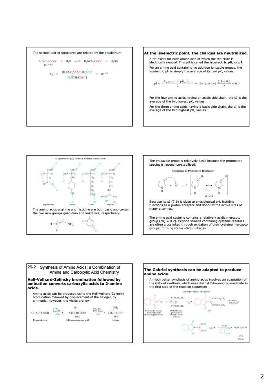
2 The second pair of structures are related by the equilibrium: At the isoelectric point, the charges are neutralized. A pH exists for each amino acid at which the structure is electrically neutral. This pH is called the isoelectric pH, or pI. For an amino acid containing no addition ionizable groups, the isoelectric pH is simply the average of its two pKa values: For the four amino acids having an acidic side chain, the pI is the average of the two lowest pKa values. For the three amino acids having a basic side chain, the pI is the average of the two highest pKa values. The amino acids arginine and histidine are both basic and contain the two new groups guanidino and imidazole, respectively: The imidazole group is relatively basic because the protonated species is resonance-stabilized: Because its pI (7.6) is close to physiological pH, histidine functions as a proton acceptor and donor at the active sites of many enzymes. The amino acid cysteine contains a relatively acidic mercapto group (pKa = 8.2). Peptide strands containing cysteine residues are often crosslinked through oxidation of their cysteine mercapto groups, forming stable –S-S- linkages. Synthesis of Amino Acids: a Combination of Amine and Carboxylic Acid Chemistry 26-2 Hell-Volhard-Zelinsky bromination followed by amination converts carboxylic acids to 2-amino acids. Amino acids can be produced using the Hell-Volhard-Zelinsky bromination followed by displacement of the halogen by ammonia, however, the yields are low. The Gabriel synthesis can be adapted to produce amino acids. A much better synthesis of amino acids involves an adaptation of the Gabriel synthesis which uses diethyl 2-bromopropanedioate in the first step of the reaction sequence:
m rolkyated.llno eacarepneparedomenvdsbythe ggn,2inariyeversbeaatoalehydestofom 0 hehelavarlation of thisc the 26-3 Synthesis of Enantiomerically Pure Amino Acids H,0 H H.O orpoiaPracticethsmthodsodousandcansuf 26-4 Peptides and Proteins:Amino Acid Oliomers and tarR5a85hpgmggmsYms:aoH ntlde hond mn thun tn o io i..fo 3
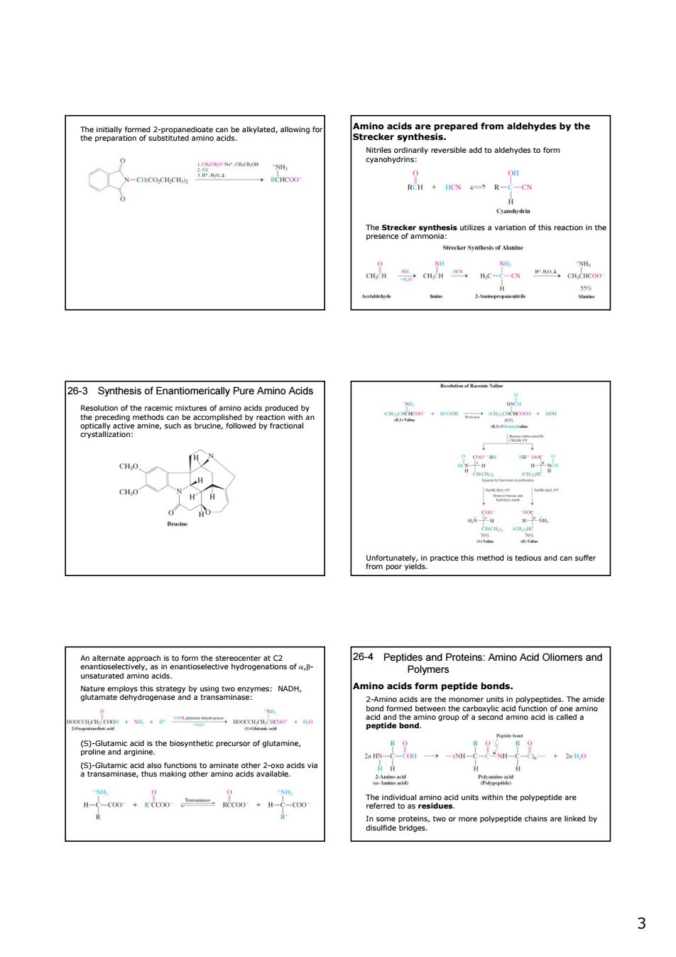
3 The initially formed 2-propanedioate can be alkylated, allowing for the preparation of substituted amino acids. Amino acids are prepared from aldehydes by the Strecker synthesis. Nitriles ordinarily reversible add to aldehydes to form cyanohydrins: The Strecker synthesis utilizes a variation of this reaction in the presence of ammonia: 26-3 Synthesis of Enantiomerically Pure Amino Acids Resolution of the racemic mixtures of amino acids produced by the preceding methods can be accomplished by reaction with an optically active amine, such as brucine, followed by fractional crystallization: Unfortunately, in practice this method is tedious and can suffer from poor yields. An alternate approach is to form the stereocenter at C2 enantioselectively, as in enantioselective hydrogenations of α,β- unsaturated amino acids. Nature employs this strategy by using two enzymes: NADH, glutamate dehydrogenase and a transaminase: (S)-Glutamic acid is the biosynthetic precursor of glutamine, proline and arginine. (S)-Glutamic acid also functions to aminate other 2-oxo acids via a transaminase, thus making other amino acids available. Peptides and Proteins: Amino Acid Oliomers and Polymers 26-4 Amino acids form peptide bonds. 2-Amino acids are the monomer units in polypeptides. The amide bond formed between the carboxylic acid function of one amino acid and the amino group of a second amino acid is called a peptide bond. The individual amino acid units within the polypeptide are referred to as residues. In some proteins, two or more polypeptide chains are linked by disulfide bridges
The peptide bond is planar and fairly rigid at room temperature. DatRertaaiuaractertzedbvthetrsoquenceod aeee2民8eont Several small peptides are physoally: eins and three disulfide prma structur 4
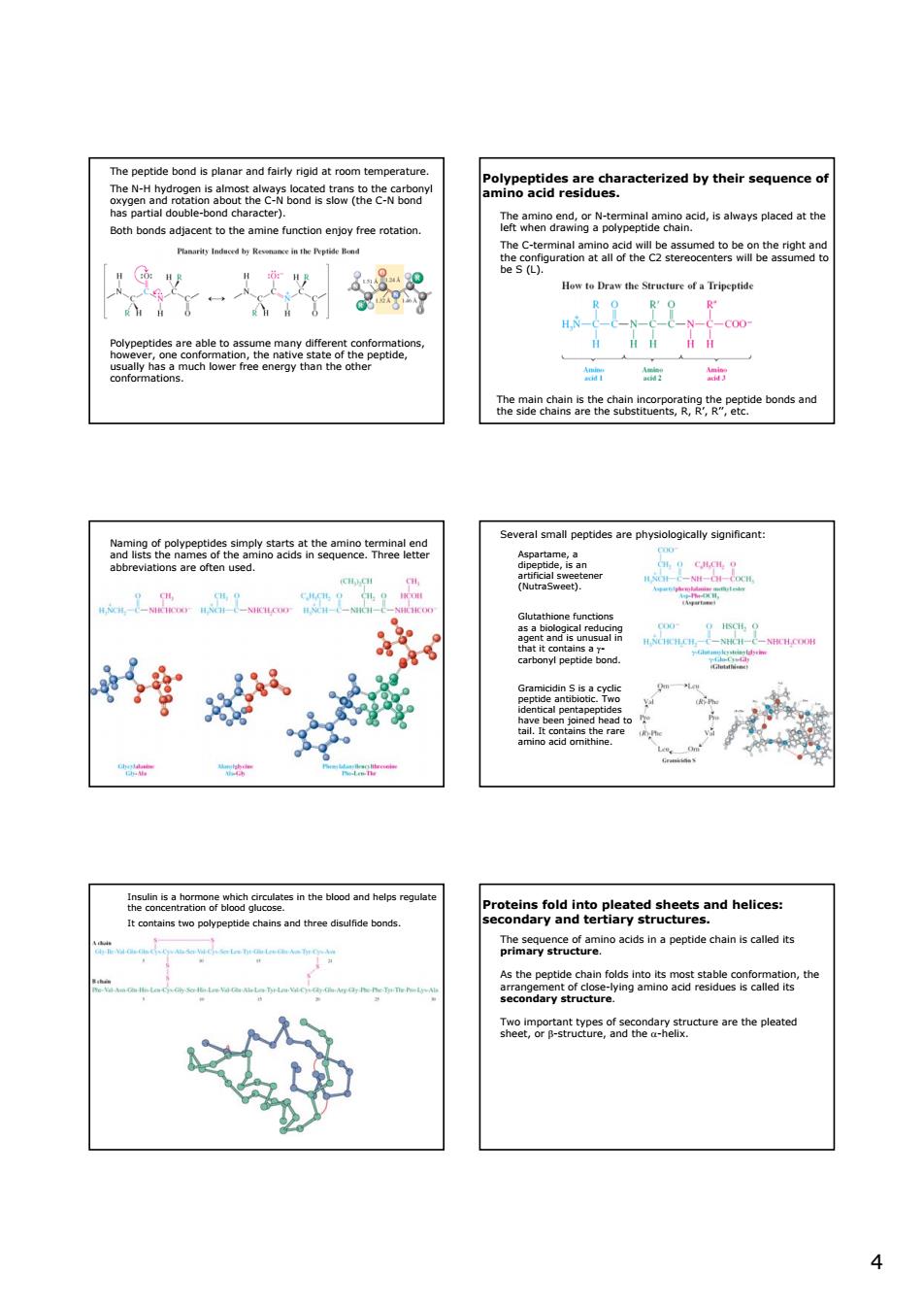
4 The peptide bond is planar and fairly rigid at room temperature. The N-H hydrogen is almost always located trans to the carbonyl oxygen and rotation about the C-N bond is slow (the C-N bond has partial double-bond character). Both bonds adjacent to the amine function enjoy free rotation. Polypeptides are able to assume many different conformations, however, one conformation, the native state of the peptide, usually has a much lower free energy than the other conformations. Polypeptides are characterized by their sequence of amino acid residues. The amino end, or N-terminal amino acid, is always placed at the left when drawing a polypeptide chain. The C-terminal amino acid will be assumed to be on the right and the configuration at all of the C2 stereocenters will be assumed to be S (L). The main chain is the chain incorporating the peptide bonds and the side chains are the substituents, R, R’, R’’, etc. Naming of polypeptides simply starts at the amino terminal end and lists the names of the amino acids in sequence. Three letter abbreviations are often used. Several small peptides are physiologically significant: Aspartame, a dipeptide, is an artificial sweetener (NutraSweet). Glutathione functions as a biological reducing agent and is unusual in that it contains a γ- carbonyl peptide bond. Gramicidin S is a cyclic peptide antibiotic. Two identical pentapeptides have been joined head to tail. It contains the rare amino acid ornithine. Insulin is a hormone which circulates in the blood and helps regulate the concentration of blood glucose. It contains two polypeptide chains and three disulfide bonds. Proteins fold into pleated sheets and helices: secondary and tertiary structures. The sequence of amino acids in a peptide chain is called its primary structure. As the peptide chain folds into its most stable conformation, the arrangement of close-lying amino acid residues is called its secondary structure. Two important types of secondary structure are the pleated sheet, or β-structure, and the α-helix

noeeheetehehegBhcabge 3名2流oe hmotcontrct 5peaeenSt6hgEsons6omearasnem - d sheets impart considerable rigidity to the syster 89phcagheaeoteesenceoftheamn tsee2Sgendnegbaarpoamseena nitonegeereeretm,.everaa-hece tre and ag 5
5 In the pleated sheet, the two chains line up with the carboxy groups of one chain opposite the amino groups of another. Additional chains may be bonded to either side to construct a “sheet” of chains connected by hydrogen bonds. This arrangement of chains can also be formed by a single polypeptide chain folding back and forth on itself several times. β-Pleated sheets impart considerable rigidity to the system. The α-helix is formed by hydrogen bonds between carbonyl groups and amino groups 3.6 amino acids apart in the amino acid sequence. The carbonyl group of one amino acid hydrogen bonds with the amino group of the amino acid four residues ahead in the sequence. Two equivalent points in adjacent turns of the helix are 5.4 Å apart. Too much charge of the same kind or the presence of the amino acid proline may disrupt secondary structure. The final overall folding of the entire polypeptide chain is called the tertiary structure of the chain. A variety of forces are involved in stabilizing the tertiary structure. •Disulfide bridges •Hydrogen bonds •London forces •Electrostatic attraction and repulsion •Micellar effects (hydrophobic effect) Pronounced folding is found in the globular proteins (chemical transport, catalysis, etc.) In fibrous proteins (myosin, fibrin, α-keratin), several α-helices are coiled together to form a superhelix. Enzymes and transport proteins fold up in such a way to produce three dimensional pockets or groves on their surfaces called active sites or binding sites. The size and shape of these sites provide a very specific fit for the intended substrate or ligand. The inner surface of an active site generally contains a specific arrangement of side chains of polar amino acids that attract functional groups on the substrate by hydrogen bonding or ionic interactions. Active sites align the functional groups on the enzyme and substrates in such a way as to promote the associated chemical reaction. A typical example of an enzyme is chymotprysin which catalyses the hydrolysis of specific peptide bonds (adjacent to phenylalanine, tyrosine or tryptophan) at physiological temperature and pH. Exposure of a protein to extremes of heat or pH usually causes denaturation, or breakdown, of the tertiary structure of a protein. In some proteins, several polypeptide chains, each with its own tertiary structure, assemble to form a larger structure called a quaternary structure